

Full-field optical coherence tomography: from micro to macro imaging
NaMeS students are invited to IPC PAS Seminar Lecture:
Egidijus Auksorius, PhD
Institut Langevin
Paris, France
Friday 20th October, 12:00 to 13:30
Assembly hall of the IPC PAS, Kasprzaka 44/52, PL-01 224 Warszawa
Abstract
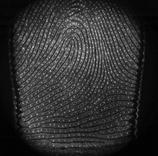
Optical coherence tomography (OCT) has become an established tool in biomedical imaging. Standard OCT is apoint-scanning interferometric technique capable of fast in vivo visualization of tissue architecture. Full-field optical coherence tomography (FF-OCT), on the other hand, uses a camera instead of a point detector and a conventional incoherent light source instead of a laser that enables parallelized detection, and thus, fast en face imaging. FF-OCT can be useful in a range of applications that require either high resolution or/and en face imaging. Thanks to its high isotropic resolution (< 1 μm in 3D), it can be used in applications that normally require the preparation of histology slides, such as in studying the enteric nervous system [1]. Spatial resolution can be traded-off for a larger field-of-view (>1 cm2) that is necessary, for example, in subsurface fingerprint imaging [2].
Images of subsurface fingerprints are of great interest in biometrics since they contain more details than the surface fingerprints and, most importantly, can be partly free of imaging artifacts caused by damage, moisture or dirt on the surface. We have built an FF-OCT subsurface fingerprint sensor based on a novel silicon camera that allowed acquisition of high quality subsurface fingerprints, and subsequently, identification of individuals with high accuracy from a single finger [3].
To increase the sensor’s performance further in terms of the signal-to-noise ration (SNR) dark-field detection can be implemented in the FF-OCT configuration [4]. It can reject spurious signal, such as specular reflections from a sample and other optical elements, that effectively allows a more efficient use of camera’s detection bandwidth. Since some of the genuine signal is also rejected in the process, a brighter light source or a configuration that utilizes the limited light budget more efficiently is thus needed. To this end, I will present a configuration that involves an asymmetric interferometer with a 10:90 beamsplitter allowing near ×4 more efficient signal detection.
The developed instrument could be used in a number of other en face deep-tissue imaging applications thanks to its high sensitivity and speed. Naturally, it could be used for imaging various skin conditions, such as cancer and other dermatological diseases.
References
[1] Coron, E., Auksorius, E., Pieretti, A., Mahé, M. M., Liu, L., Steiger, C., ... & Goldstein, A. M. (2012). Full-‐‑field optical coherence microscopy is a novel technique for imaging enteric ganglia in the gastrointestinal tract. Neurogastroenterology & Motility, 24(12).
[2] Auksorius, E., & Boccara, A. C. (2015). Fingerprint imaging from the inside of a finger with full-field optical coherence tomography. Biomedical optics express, 6(11), 4465-4471.
[3] Auksorius, E., & Boccara, A. C. (2017). Fast subsurface fingerprint imaging with full-field optical coherence tomography system equipped with a silicon camera. Journal of Biomedical Optics, 22(9), 096002.
[4] Auksorius, E., & Boccara, A. C. (2015). Dark-field full-field optical coherence tomography. Optics letters, 40(14), 3272-3275.
